| Project related papers |
ACS Photonics, 2018, 5 (8), pp 3372-3378, DOI: 10.1021/acsphotonics.8b00636
- Corresponding ORDP dataset: DOI: 10.13140/RG.2.2.19079.65441
Stefan Mastel, Alexander A. Govyadinov, Curdin Maissen, Andrey Chuvilin, Andreas Berger, and Rainer Hillenbrand
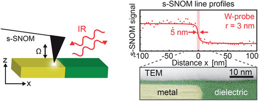
Abstract: Scattering-type scanning near-field optical microscopy (s-SNOM) allows for nanoscale-resolved Infrared (IR) and Terahertz (THz) imaging, and thus has manifold applications ranging from materials to biosciences. However, a quantitatively accurate understanding of image contrast formation at materials boundaries, and thus spatial resolution is a surprisingly unexplored terrain. Here we introduce the read/write head of a commercial hard disk drive (HDD) as a most suitable test sample for fundamental studies, given its well-defined sharp material boundaries perpendicular to its ultrasmooth surface. We obtain unprecedented and unexpected insights into the s-SNOM image formation process, free of topography-induced contrasts that often mask and artificially modify the pure near-field optical contrast. Across metal-dielectric boundaries, we observe non-point-symmetric line profiles for both IR and THz illumination, which are fully corroborated by numerical simulations. We explain our findings by a sample-dependent confinement and screening of the near fields at the tip apex, which will be of crucial importance for an accurate understanding and proper interpretation of high-resolution s-SNOM images of nanocomposite materials. We also demonstrate that with ultrasharp tungsten tips the apparent width (resolution) of sharp material boundaries can be reduced to about 5 nm.
Probes for Ultrasensitive THz Nanoscopy
ACS Photonics2019, 6, 5, 1279-1288 DOI: 10.1021/acsphotonics.9b00324
- Corresponding ORDP dataset: DOI: 10.13140/RG.2.2.28694.50246
Curdin Maissen, Shu Chen, Elizaveta Nikulina, Alexander Govyadinov and Rainer Hillenbrand
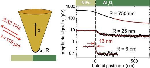
Abstract: Scattering-type scanning near-field microscopy (s-SNOM) at terahertz (THz) frequencies could become a highly valuable tool for studying a variety of phenomena of both fundamental and applied interest, including mobile carrier excitations or phase transitions in 2D materials or exotic conductors. Applications, however, are strongly challenged by the limited signal-to-noise ratio. One major reason is that standard atomic force microscope (AFM) tips, which have made s-SNOM a highly practical and rapidly emerging tool, provide weak scattering efficiencies at THz frequencies. Here, we report a combined experimental and theoretical study of commercial and custom-made AFM tips of different apex diameter and length, in order to understand signal formation in THz s-SNOM and to provide insights for tip optimization. Contrary to common beliefs, we find that AFM tips with large (micrometer-scale) apex diameter can enhance s-SNOM signals by more than 1 order of magnitude, while still offering a spatial resolution in the 100 nm range at a wavelength of λ = 119 μm. On the other hand, exploiting the increase of s-SNOM signals with tip length, we succeeded in sub-15 nm (<λ/8000) resolved THz imaging employing a tungsten tip with 6 nm apex radius. We explain our findings and provide novel insights into s-SNOM via rigorous numerical modeling of the near-field scattering process. Our findings will be of critical importance for pushing THz nanoscopy to its ultimate limits regarding sensitivity and spatial resolution.
Plasmonic Antennas with Electric, Magnetic, and Electromagnetic Hot Spots Based on Babinet’s Principle
Phys. Rev. Applied 13, 054045 – Published 19 May 2020 DOI: 10.1103/PhysRevApplied.13.054045
- Corresponding ORDP dataset: DOI: 10.13140/RG.2.2.18865.81766
Martin Hrtoň, Andrea Konečná, Michal Horák, Tomáš Šikola, and Vlastimil Křápek
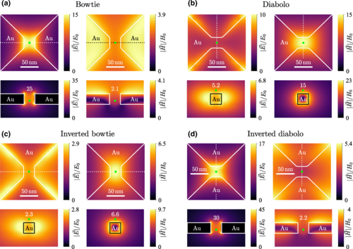
Abstract: We theoretically study plasmonic antennas featuring areas of extremely concentrated electric or magnetic field, known as hot spots. We combine two types of electric-magnetic complementarity to increase the degree of freedom for the design of the antennas: bowtie and diabolo duality and Babinet’s principle. We evaluate the figures of merit for different plasmon-enhanced optical spectroscopy methods and optical trapping: field enhancement, decay rate enhancement, quality factor of the plasmon resonances, and trapping potential depth. The role of Babinet’s principle in interchanging electric and magnetic field hot spots and its consequences for practical antenna design are discussed. In particular, diabolo antennas exhibit slightly better performance than bowties in terms of larger field enhancement and larger Q factor. For specific resonance frequency, diabolo antennas are considerably smaller than bowties, which makes them favorable for the integration into more complex devices but also makes their fabrication more demanding in terms of spatial resolution. Finally, we propose a Babinet-type dimer antenna featuring electromagnetic hot spot with both the electric and magnetic field components treated on an equal footing.
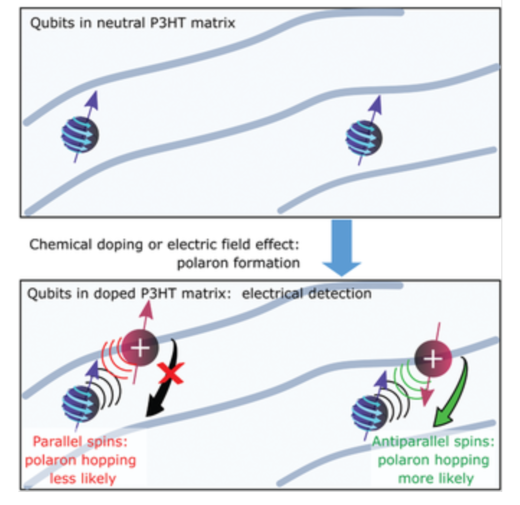
Hybrid Spintronic Materials from Conducting Molecular Quantum Bits
Adv. Funct. Mater. 2020, 20006882, DOI: 10.1002/adfm.202006882
Michal Kern, Lorenzo Tesi, David Neusser, Nadine Rußegger, Mario Winkler, Alexander Allgaier, Yannic M. Gross, Stefan Bechler, Hannes S. Funk, Li‐Te Chang, Jörg Schulze, Sabine Ludwigs, and Joris van Slageren
Abstract: Hybrid materials consisting of organic semiconductors and molecular quantum bits promise to provide a novel platform for quantum spintronic applications. However, investigations of such materials, elucidating both the electrical and quantum dynamical properties of the same material have never been reported. Here the preparation of hybrid materials consisting of conducting polymers and molecular quantum bits is reported. Organic field‐effect transistor measurements demonstrate that the favorable electrical properties are preserved in the presence of the qubits. Chemical doping introduces charge carriers into the material, and variable‐temperature charge transport measurements reveal the existence of mobile charge carriers at temperatures as low as 15 K. Importantly, quantum coherence of the qubit is shown to be preserved up to temperatures of at least 30 K, that is, in the presence of mobile charge carriers. These results pave the way for employing such hybrid materials in novel molecular quantum spintronic architectures.
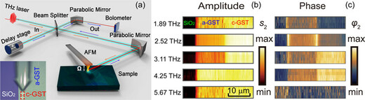
Terahertz Nanoimaging and Nanospectroscopy of Chalcogenide Phase-Change Materials
ACS Photonics 2020, 7, 12, 3499–3506 Publication Date:November 30, 2020 DOI:10.1021/acsphotonics.0c01541
by Chao Chen, Shu Chen, Ricardo P.S.M. Lobo, Carlos Maciel-Escudero, Martin Lewin, Thomas Taubner, Wei Xiong, Ming Xu, Xinliang Zhang, Xiangshui Miao, Peining Li, and Rainer Hillenbrand
Abstract: Chalcogenide phase-change materials (PCMs) exhibit optical phonons at terahertz (THz) frequencies, which can be used for studying basic properties of the phase transition and which lead to a strong dielectric contrast that could be exploited for THz photonics applications. Here, we demonstrate that the phonons of PCMs can be studied by frequency-tunable THz scattering-type scanning near-field optical microscopy (s-SNOM). Specifically, we perform spectroscopic THz nanoimaging of a PCM sample comprising amorphous and crystalline phases. We observe phonon signatures, yielding strong s-SNOM signals and, most important, clear spectral differences between the amorphous and crystalline PCM, which allows for distinguishing the PCM phases with high confidence on the nanoscale. We also found that the spectral signature can be enhanced, regarding both signal strength and spectral contrast, by increasing the radius of the probing tip. From a general perspective, our results establish THz s-SNOM for nanoscale structural and chemical mapping based on local phonon spectroscopy.
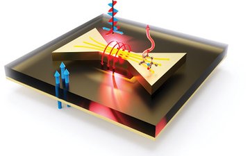
by Lorenzo Tesi, Dominik Bloos, Martin Hrtoň, Adam Beneš, Mario Hentschel, Michal Kern, Alisa Leavesley, Rainer Hillenbrand, Vlastimil Křápek, Tomáš Šikola, and Joris van Slageren
Abstract: Nanoscale magnetic systems play a decisive role in areas ranging from biology to spintronics. Although, in principle, THz electron paramagnetic resonance (EPR) provides high-resolution access to their properties, lack of sensitivity has precluded realizing this potential. To resolve this issue, the principle of plasmonic enhancement of electromagnetic fields that is used in electric dipole spectroscopies with great success is exploited, and a new type of resonators for the enhancement of THz magnetic fields in a microscopic volume is proposed. A resonator composed of an array of diabolo antennas with a back-reflecting mirror is designed and fabricated. Simulations and THz EPR measurements demonstrate a 30-fold signal increase for thin film samples. This enhancement factor increases to a theoretical value of 7500 for samples confined to the active region of the antennas. These findings open the door to the elucidation of fundamental processes in nanoscale samples, including junctions in spintronic devices or biological membranes.